

Key Takeaways
Brain imaging techniques employed for diagnosing depression encompass Magnetic Resonance Imaging (MRI), Functional MRI (fMRI), and Positron Emission Tomography (PET). Each serves a distinct role, revealing various aspects of brain activity and structure. MRI zeroes in on structural anomalies, while fMRI gauges blood flow to pinpoint active areas. PET, meanwhile, highlights metabolic activity that can be linked to depression.
Identifiable neural patterns from these scans can shed light on different forms of depression and inform treatment decisions. For instance, major depressive disorder typically showcases decreased activity in certain brain regions, whereas individuals with bipolar disorder may display marked differences in brain activity.
Integrating findings from brain imaging with traditional assessments paves the way for tailored treatment plans, moving away from a standardized method. This combination has the potential to enhance therapeutic outcomes by aligning treatments with observed brain activity, particularly in regions such as the amygdala.
Noteworthy applications of brain scans have demonstrated increased success rates in difficult-to-treat cases. A case in point involves personalized strategies that blend psychotherapy with neurofeedback, showing promise for patients grappling with atypical depression.
Grasping the connection between brain activity and symptoms of depression is essential. Key brain regions like the prefrontal cortex and amygdala are crucial for emotional regulation and fear processing, thus impacting therapeutic choices.
By merging imaging data with clinical evaluations, the accuracy of diagnosis and treatment strategies is significantly improved. This collaborative approach benefits both clinicians and patients, ensuring that treatment plans are responsive to individual brain activity patterns.
Individuals preparing for a brain scan should anticipate an initial consultation, adhere to specific scan preparation guidelines, and participate in a post-scan discussion to explore findings. Such a process aims to ease anxiety and foster comprehension of the results and subsequent treatment options.
Choosing the Right Brain Scan for Depression Diagnosis
Types of Brain Imaging Techniques Explained
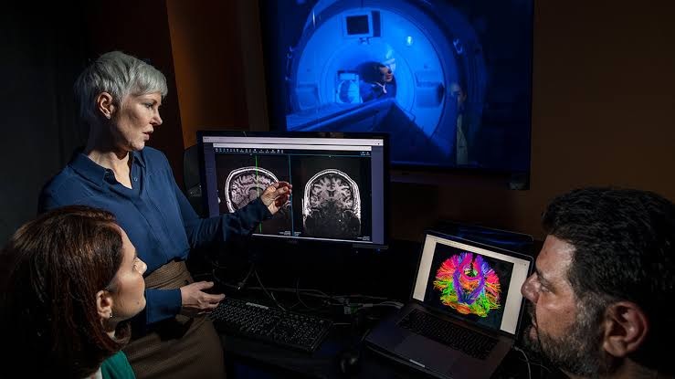
When diagnosing depression, grasping the array of brain imaging techniques available is vital. Multiple methodologies exist, each with its unique strengths. Among them, professionals often highlight three main types: Magnetic Resonance Imaging (MRI), Functional MRI (fMRI), and Positron Emission Tomography (PET). Each technique delivers distinct insights regarding brain function.
An MRI excels at producing comprehensive images of brain structures. It’s particularly effective for identifying physical abnormalities like lesions or tumors. Its non-invasive nature establishes MRI as a go-to choice for initial assessments. However, for diagnosing depression, relying solely on structural details may fall short.
Contrastingly, fMRI monitors brain activity by detecting alterations associated with blood flow. This technique offers professionals a window into which brain areas become active during specific thoughts or emotions. In other words, it’s akin to viewing the brain in real time.
PET scans, albeit not as commonly employed as MRI and fMRI, evaluate metabolic activity. By identifying regions of the brain with heightened glucose consumption, they can signal potential imbalances related to depression.
- Magnetic Resonance Imaging (MRI): Primarily identifies structural issues.
- Functional MRI (fMRI): Reveals brain activity and measures blood flow.
- Positron Emission Tomography (PET): Analyzes metabolic function and activity.
How Scans Reveal Unique Depression Patterns
Experts emphasize that the distinctive patterns uncovered through brain scans provide crucial insights into depression. A brain scan for depression frequently reveals unique neural pathways and connections associated with various types of depression.
For example, studies indicate that individuals diagnosed with major depressive disorder often show decreased activity in areas such as the prefrontal cortex or the anterior cingulate cortex. These reductions can signify challenges in emotional regulation and decision-making. Yet, there’s always more to consider.
People experiencing bipolar disorder may demonstrate variations in brain activity divergent from those with unipolar depression. These differences underscore the necessity of customizing treatment to each individual’s specific neurobiological framework.
Understanding these neural signatures is paramount. By leveraging brain imaging, healthcare professionals can determine the types of depression patients are facing, ultimately leading to improved treatment approaches.
The Role of Brain Imaging in Personalized Treatment Plans
Tailoring Treatment Based on Imaging Results
In crafting treatment plans, professionals stress the importance of integrating imaging results with conventional assessments. Brain scans allow practitioners to shift away from cookie-cutter treatment models. Instead, the focus is on personalized and targeted strategies shaped by individual brain activity patterns.
This pivot toward personalized medicine can significantly enhance outcomes. For instance, if an expert identifies elevated activity in the amygdala—a region linked to fear responses and emotional processing—the treatment approach may adapt. Aligning therapeutic options with specific brain activity can lead to better patient responses.
- Medications: Adjusting antidepressant regimens based on insights obtained from imaging.
- Therapeutic Approaches: Customizing psychotherapy methods to complement discovered patterns.
- Lifestyle Changes: Adopting comprehensive strategies that resonate with findings.
However, it’s essential to bear in mind that imaging is just one part of a larger treatment puzzle. When thoughtfully integrated, it deepens understanding of how patients can journey along their path to healing.
Real-World Examples of Successful Outcomes
Theoretical discussions can be fascinating, but real-world scenarios often tell a more compelling story. Numerous success stories highlight how brain scans can result in breakthrough improvements, particularly for treatment-resistant cases. Consider a patient diagnosed with atypical depression; imaging exposed unusual activity in areas tied to mood regulation, prompting professionals to implement a blend of psychotherapy and neurofeedback techniques.
This approach led to notable progress over time, with considerable reductions in symptom severity. Another example features an individual with a history of recurrent depressive episodes. Here, brain scans unveiled abnormal brain connectivity, guiding treatment towards a specific regimen that utilized medications influenced by imaging data.
“The brain is an incredibly complex organ. Personalized imaging insights allow us to work through treatment options like never before.” – Leading Neuropsychiatrist
The overarching narrative is compelling: customizing treatments based on imaging findings fosters more meaningful advances. Patients often discover renewed hope where traditional methods may have missed the mark.
Understanding the Science Behind Brain Scans and Depression
How Brain Activity Correlates with Depression Symptoms
Professionals have achieved impressive progress in revealing the connection between brain activity and symptoms of depression. By examining neural patterns, experts unravel a comprehensive narrative about how connectivity impacts emotional states.
Key brain regions, such as the prefrontal cortex—critical for executive functions—often demonstrate reduced activity in those with depression. Such inactivity is closely tied to diminished motivation and cognitive sluggishness. However, this is just scratching the surface.
The interplay between brain activity and depression symptoms reveals a growing complexity. Certain neural pathways may become overactive while others fall short, resulting in confusing emotional responses. For instance, during a depressive episode, individuals may experience an overactive amygdala, which amplifies feelings of fear and anxiety.
- Prefrontal Cortex: Associated with decision-making and emotional regulation.
- Amygdala: Central to processing fear and anxiety.
- Hippocampus: Important for memory and emotional responses.
Exploring these connections can illuminate appropriate therapeutic avenues, revitalizing focus on effective treatment strategies.
Integrating Imaging Data with Clinical Assessments
Successful diagnosis and treatment heavily rely on the effective merging of imaging data with clinical evaluations. Standard assessment methods might overlook critical insights, but combining these results offers a comprehensive view of each patient’s mental health landscape.
Clinical assessments deliver context concerning a patient’s lived experiences, whereas imaging provides an understanding of the underlying brain activity. For instance, while a therapist may recognize symptoms of chronic sadness, the combination with imaging data could reveal identifiable neural patterns affecting those sentiments. This collaborative effort enables the creation of far more effective treatment plans.
- Patient History: Crucial for contextualizing imaging results.
- Symptom Tracking: Assists in identifying trends over time.
- Regular Assessments: Ensures treatment plans stay aligned with shifts in brain activity.
This integrated strategy not only guides clinicians but also empowers patients to engage actively in their treatment journey.
What to Expect During the Brain Imaging Process
Preparing for Your Scan: A Step-by-Step Guide
Grasping what to expect during the brain imaging process can greatly assuage anxiety. Many individuals approach brain scans with trepidation, often based on misunderstandings. Here’s a straightforward step-by-step outline.
- Initial Consultation: Expect an in-depth conversation with a medical professional to assess the necessity of the scan.
- Prepping for the Scan: Remove all metal possessions; change into a gown if needed.
- Scan Procedure: Lie comfortably on the scanning table, making any necessary adjustments.
- Post-Scan: Participate in a follow-up consultation to discuss the findings.
Many people question the potential for discomfort. In practice, most find the experience quite manageable—generally, patients aren’t burdened with invasive equipment during the scan.
Post-Scan Insights and Next Steps
After completing the brain scans, what does the journey ahead look like? Understanding the results is crucial for determining subsequent actions. For many, a comprehensive analysis from a specialist enhances clarity regarding findings.
In the follow-up consultation, results will be explained in detail. Often, these insights trigger discussions centered on personalized treatment pathways. Patients should feel encouraged to ask questions and review treatment options grounded in their distinctive brain activity patterns.
Though every patient’s path is unique, engaging actively in this dialogue fosters an important bond between patient and provider. After all, knowledge is empowering.
“The insights gained from brain scans illuminate unique pathways to healing.” – Leading Neuropsychiatrist
Each step along the road to recovery is bolstered by enhanced understanding of the mind’s intricate interrelations. Such dialogue is fundamental in establishing a robust basis for healing.
FAQ
What is the role of neurofeedback in treating depression?
Neurofeedback represents a cutting-edge strategy that trains individuals to modify their brain activity. By offering real-time feedback, this method aids patients in developing self-regulation skills, often resulting in significant symptom improvements for many dealing with various forms of depression.
Are there specific symptoms that brain imaging can track over time?
Indeed, brain imaging can monitor shifts in brain activity connected to particular symptoms. For instance, fluctuations in amygdala activity may correlate with heightened anxiety, while variations in the prefrontal cortex’s engagement might reflect changes in motivation or mood.
How do lifestyle factors influence depression diagnoses?
Lifestyle elements, including nutrition, physical activity, and sleep, can profoundly influence the intensity of depression symptoms and overall brain health. Incorporating this understanding alongside imaging findings allows professionals to personalize recommendations that resonate with a patient’s needs, thus enhancing treatment effectiveness.
Can brain imaging be combined with other diagnostic tools?
Absolutely. Merging brain imaging with psychological evaluations and standardized questionnaires often provides a more nuanced view of a patient’s mental health. This approach paves the way for well-informed clinical decisions and customized treatment strategies.
Is there an age limit for patients undergoing brain scans for depression?
No strict age limits govern the practice, though younger patients typically undergo supplementary evaluations to confirm the scanning necessity. Tailoring the scanning process to a patient’s age and development is vital for accurate assessment and treatment planning.
What advancements are expected in brain imaging technology for depression?
Future developments may feature improved imaging techniques that yield higher resolution and deeper insights into real-time brain functions. Innovations, such as portable imaging devices, could expand access to these diagnostic tools, making them more readily available to a wide range of patient populations.
How effective is brain imaging for uncovering treatment-resistant depression?
Brain imaging has shown significant potential in identifying underlying neurobiological factors contributing to treatment resistance. By grasping these patterns, clinicians can tailor interventions that help patients find relief when traditional treatments fall short.
What can patients do to prepare mentally for a brain scan?
Preparing mentally involves familiarizing oneself with the scan’s purpose and procedure. Engaging in discussions about any concerns with a healthcare provider can help patients set realistic expectations and ensure they feel supported throughout the experience.
Useful Resources
- National Institute of Mental Health (NIMH)
- American Psychological Association (APA)
- Psychology Today
- National Center for Biotechnology Information (NCBI)
- gov
- American Psychiatric Association
- Society for Neuroscience – BrainFacts
- APA Topic Page on Depression
- Mayo Clinic
- Healthline
